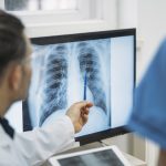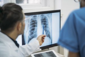
A chest X-ray is one of the most common radiology tests in modern medicine. It offers a quick, non-invasive way to visualize the structures inside the chest — including the lungs, heart, bones, and major blood vessels. Whether you’re experiencing breathing problems, chest pain, or undergoing routine screening, a chest X-ray can provide vital clues about your health.
This article dives into why doctors order chest X-rays, what these images reveal, and how you can best prepare for the procedure.
What Is a Chest X-Ray?
A chest X-ray is a radiographic image of the chest area using a small dose of ionizing radiation to create pictures of the organs and structures inside the chest. It is a quick and painless test that usually takes just a few minutes to complete.
It typically provides images of:
- Lungs
- Heart
- Ribs
- Diaphragm
- Spine (upper portion)
- Chest X-rays can be frontal (posteroanterior) or lateral (side view) — sometimes both are taken for better clarity.
Why Is a Chest X-Ray Ordered?
Doctors may recommend a chest X-ray for various diagnostic, monitoring, or screening purposes. Common reasons include:
1. Detecting Respiratory Conditions
A chest X-ray can help identify:
- Pneumonia
- Tuberculosis (TB)
- Chronic Obstructive Pulmonary Disease (COPD)
- Lung cancer
- Pulmonary fibrosis
- Collapsed lung (pneumothorax)
These conditions often show up as abnormal shadows, fluid accumulation, or changes in lung size and shape.
2. Evaluating Heart-Related Problems
While not as detailed as an echocardiogram or CT scan, a chest X-ray can give clues about:
- Enlarged heart (cardiomegaly)
- Heart failure
- Fluid around the heart (pericardial effusion)
- An enlarged silhouette on the X-ray may indicate an underlying cardiac issue.
3. Investigating Chest Pain or Trauma
For individuals with chest pain, injury, or shortness of breath, a chest X-ray can help rule out:
- Broken ribs
- Fluid in or around the lungs (pleural effusion)
- Air leak into the chest cavity
- Tumors or masses
4. Monitoring Ongoing Medical Conditions
Doctors may use chest X-rays to monitor:
- Recovery from pneumonia or TB
- Response to cancer treatment
- Progression of chronic lung diseases
5. Pre-Surgical Evaluation
Some surgeries require a chest X-ray as part of the preoperative assessment to rule out active infections or underlying lung issues.
6. Screening for Occupational Lung Diseases
Workers exposed to dust, asbestos, or chemicals may undergo periodic chest X-rays to monitor lung health and catch early signs of occupational diseases like asbestosis or silicosis.
What Can a Chest X-Ray Show?
A chest X-ray offers a visual snapshot that can reveal:
- Lung Abnormalities
- Infections: Cloudy or opaque areas may suggest pneumonia or TB.
- Masses or Nodules: Possible signs of lung cancer or benign growths.
- Collapsed Lung: Appears as a loss of volume on one side.
- Scarring or Fibrosis: Indicates chronic damage or past infections.
- Heart and Blood Vessel Changes
- Enlarged Heart: A sign of congestive heart failure or cardiomyopathy.
- Aortic Aneurysm: Can sometimes be detected as a widening of the aorta.
- Fluid Overload: Suggests heart failure or kidney dysfunction.
- Bone and Diaphragm Issues
- Fractured Ribs or Spine Deformities
- Abnormal Diaphragm Position: Can signal nerve damage or abdominal issues pushing on the chest cavity.
How to Prepare for a Chest X-Ray?
Chest X-rays require minimal preparation, but a few simple steps can help ensure accurate results:
Before the Test
- Wear loose, comfortable clothing without metal buttons or zippers.
- Remove jewelry, eyeglasses, or piercings that may interfere with the image.
- You may be asked to change into a hospital gown.
Inform the Technician If:
- You are pregnant or may be pregnant. Though the radiation exposure is low, extra precautions or alternative tests may be advised.
- You have had a recent X-ray or CT scan, as prior images might be useful for comparison.
During the Test
- You’ll be asked to stand against the X-ray machine or lie down on a table.
- A technician may ask you to hold your breath for a few seconds during image capture to prevent blurring.
- The process usually takes 5 to 10 minutes.
Are There Any Risks?
Chest X-rays are generally safe. The amount of radiation used is very low and unlikely to cause harm in healthy adults.
However:
- Repeated exposure to radiation should be avoided when unnecessary.
- Pregnant women should discuss alternatives unless the benefits outweigh the risks.
After the Chest X-Ray
There is no recovery time — you can resume normal activities right away. The images will be reviewed by a radiologist, and your doctor will explain the results.
Your results might show:
- Normal findings – No further action required.
- Abnormal but non-serious conditions – Such as minor infections.
- Significant findings – Which may require further tests like a CT scan, MRI, or biopsy.
When Should You Be Concerned About Chest Symptoms?
See a doctor if you experience:
- Persistent cough or wheezing
- Chest pain not related to injury
- Shortness of breath
- Coughing up blood
- Unexplained fever, fatigue, or weight loss
- A chest X-ray can be the first step in identifying the cause.
Conclusion
A chest X-ray helps doctors quickly assess conditions affecting the lungs, heart, and chest wall. It is safe, fast, and widely used for both diagnosis and follow-up. Whether you’re dealing with a respiratory infection or undergoing routine screening, understanding the purpose and process of a chest X-ray can help ease anxiety and improve communication with your healthcare provider.
Always follow your doctor’s advice regarding follow-up testing and treatment based on your chest X-ray findings.