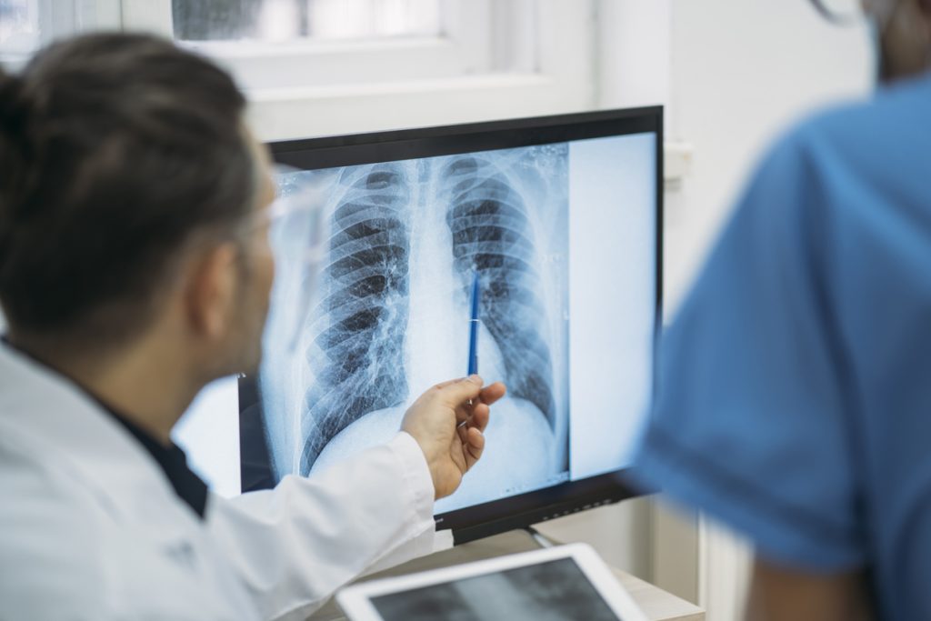
What is a Chest X-Ray?
A chest x-ray is a type of medical imaging procedure that uses x-rays to look at structures and organs of the chest. It helps to see how well the lungs and heart are working.
The following are diagnosed by a chest x-ray:
- Heart
- Lungs
- Bronchi
- Aorta
- Pulmonary Arteries
- Middle Chest Area
- Bones of the chest
What symptoms require the need for a Chest X-Ray?
A chest x-ray is ordered to diagnose issues that cause symptoms in the heart or lungs. Some of the symptoms include
- Difficulty in breathing: This could signal lung conditions like pneumonia, asthma, fluid buildup, or heart failure.
- Chronic cough: A persistent cough may indicate infections, lung disease, or even tumors.
- Chest pain: This could be related to heart problems, lung infections, collapsed lungs, or rib injuries.
- Fever with other signs of infection: A high fever along with a cough or chest discomfort might point to pneumonia or other respiratory infections.
What conditions can a Chest X-Ray diagnose?
A Chest X-Ray can be ordered to diagnose or monitor certain health conditions, including
- Pneumonia
- Lung cancer
- Tuberculosis
- Ribcage Injuries
- Chronic Obstructive Pulmonary Disease (COPD)
- Congestive Heart Failure
How does a Chest X-Ray work?
X-Rays are a form of radiation like radio waves or light. They travel through most objects, including the body. The radiologist aims the x-ray beam at the area of interest. The x-ray machine produces a burst of radiation that passes through the body. The radiation records an image on photographic film or a special detector.
Different parts of the body absorb x-rays in varying degrees. Dense bones absorb much of the radiation, while soft tissues, including the muscle, fat, and organs, allow more of the radiation to pass through them. As a result, bones appear white in x-rays, soft tissues come up in shades of grey, and air in black.
On a chest x-ray, the ribs and spine absorb much of the radiation and appear white or light grey in the image. Lung tissues absorb little radiation and will appear dark on the image.
How to prepare for a Chest X- Ray?
Chest x-rays require little to no preparation. The way this procedure is performed depends on the individual’s condition and the healthcare provider’s practices:
Generally, it follows this process:
- The individual will be asked to remove any clothing, jewelry, or metal objects that could interfere with the X-ray image.
- They should wear loose, comfortable clothing or maybe be given a hospital gown to wear.
- Depending on the type of image required, the individual may be asked to stand, sit, or lie down.
For a front view:
- The individual will stand or sit in front of the X-ray plate.
- They will be instructed to roll their shoulders forward, take a deep breath, and hold it while the image is captured.
- If they are unable to hold their breath, the radiologist will time the image with their natural breathing.
- For a side view:
- The individual will turn to one side and raise their arms above their head.
- They will take a deep breath and hold it as the X-ray is taken.
- The individual must remain completely still during the X-ray to prevent image blurring.
- The technologist will step behind a protective window or shield while capturing the images.
- The entire procedure is quick, painless, and typically takes only a few minutes.
During the procedure, one should try to remain extremely still while holding their breath. Any movement, even breathing in and out, can blur the resultant image.
What are the risks of a Chest X-Ray?
An individual may be concerned about radiation exposure from chest X-rays, especially if the procedure is performed regularly. However, the amount of radiation from a chest X-ray is low, often even lower than the natural background radiation encountered in daily life.
While the benefits of a chest X-ray typically outweigh the risks, a protective apron may be provided if multiple images are needed. If the individual is pregnant or suspects they might be pregnant, it is important that they inform the radiologist. In such cases, the procedure can be adjusted to protect the abdomen from radiation exposure.
When should I know the results of a Chest X-Ray?
The results of a chest X-ray are usually available within a day or two, depending on the facility and the urgency of the case. In many hospitals or clinics, a radiologist will examine the X-ray and send a report to the referring physician. If the X-ray is done in an emergency or urgent care setting, the results may be available within a few hours. One’s healthcare provider will then review the findings and discuss any next steps or further tests, if needed.
What do the results of a Chest X-Ray mean?
The results of a chest x-ray usually mean:
- A normal chest x-ray shows clear lungs, a healthy heart, and a clearly outlined chest cavity. There are no visible nodules, tumors, or masses.
- An abnormal chest x-ray can highlight unusual characteristics like an enlarged heart, fluid buildup in the lungs, cysts, broken ribs, masses, and other irregularities.
If one gets abnormal results, then he or she may be recommended additional tests like a CT Scan or PET Scan for further investigation.
FAQs
1. Why are chest x-rays ordered?
Chest X-rays are used to detect lung conditions, heart problems, infections, broken ribs, or abnormalities in the chest area.
2. How do I prepare for a chest x-ray?
No special preparation is needed. Just remove any jewelry or metal objects and wear a hospital gown if required.