What is mammography?
Mammography is an imaging examination of breast tissues with low-dose x-rays for any abnormalities, particularly breast cancer. The image produced by this procedure is called a mammogram, which helps detect changes in the breast, often before any physical symptoms appear.
There are two main types of mammograms:
- Screening Mammogram: This kind of mammogram is performed routinely for females without symptoms to detect hidden issues.
- Diagnostic Mammogram: This type of mammogram is examined when there are visible signs of any lumps, pain, or nipple discharge.
What symptoms indicate the need for mammography?
Mammography may be recommended if an individual experiences any of the following symptoms:
- A lump or thickening in the breast or underarm area
- Breast pain or tenderness that is persistent or unusual
- Nipple discharge, especially if it’s bloody or occurs without squeezing
- Changes in breast shape or size not related to one’s menstrual cycle
- Skin changes on the breast, such as dimpling, redness, or puckering
- Nipple changes, including inversion, scaling, or crusting
- Swelling of a part or whole of the breast
- Unexplained bruising or warmth in the breast
What conditions can mammography be diagnosed?
Mammography can detect breast cancers, benign tumors, and cysts.
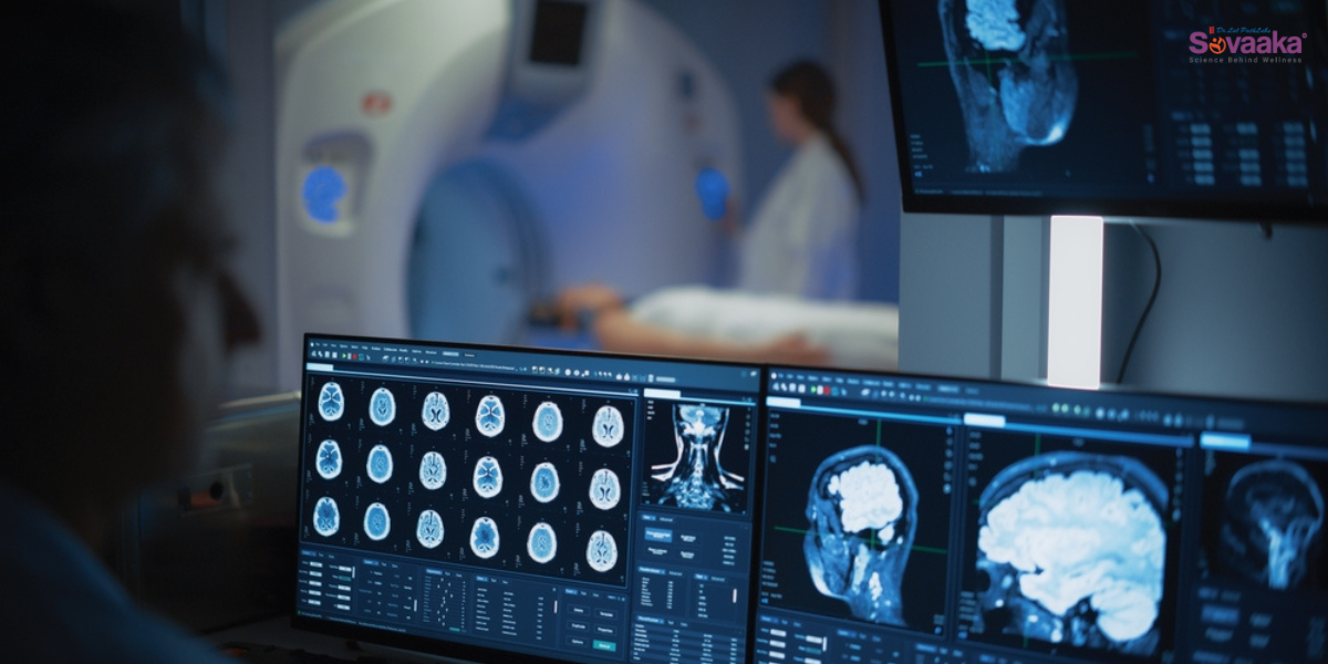
How does mammography work?
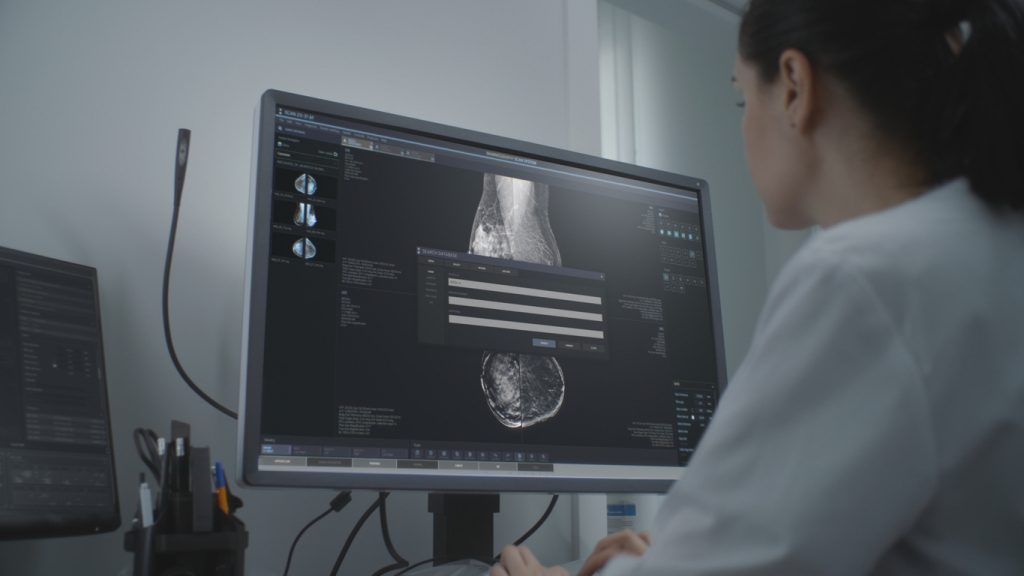
Mammography works by using low-dose x-rays to take detailed images of the inside of the breast. During the procedure, each breast is placed between two flat plates on a machine. These plates gently press the breast to spread out the tissue. This compression is important as it helps to get a clearer image and reduces the amount of radiation needed.
Once the breast is compressed, the machine takes x-ray images from different angles. These images, called mammograms, show the structure of the breast, including fats, glands, ducts, and any unusual lumps or changes. The radiologist will look at these images to check for abnormalities, such as masses, cysts, calcifications, or signs of cancer.
What preparations are required for mammography?
Before undergoing mammography, the following preparations and precautions can help ensure accurate results and a more comfortable experience:
- If an individual is breastfeeding, pregnant, or thinks they may be pregnant, then they have to report to their healthcare provider. They may recommend the best time to get a mammogram.
- One should try not to schedule the procedure a week before or during the week of menstrual period, as the breasts tend to be tender during this time, making it uncomfortable to examine.
- If one has any kind of breast implants or has recently been vaccinated, it is crucial to inform the healthcare provider.
What will happen during a mammogram?
On the day of the mammogram, an individual is advised to abide by these guidelines:
- The individual should follow their normal daily routine, including eating meals and taking prescribed medications on time.
- They should avoid using deodorant, perfume, lotion, or body powder on the day of the examination, as these substances can interfere with the accuracy of the X-ray images.
- During the procedure, the individual will be asked to undress from the waist up, and a medical gown or drape will be provided for modesty.
- The individual will stand in front of the mammography machine. The radiologist will ask them to remove one breast at a time from the gown for imaging.
- Each breast will be placed on a breast support plate, and a plastic paddle will gently compress it to spread the tissue evenly.
- Mild discomfort may be experienced during compression. If the individual finds the pressure too uncomfortable, they should inform the radiologist so that adjustments can be made.
- X-ray images will be taken while the breast is compressed.
- After the images have been captured, the examination will be complete, and the individual can get dressed.
How long does mammography take?
Screening mammograms take about 15 to 20 minutes. Diagnostic mammograms may take longer due to the radiologist needing extra images for a deeper evaluation.
What are the risks associated with mammography?
Mammography, while generally safe, may involve the following risks:
- Radiation Exposure: Mammography uses low-dose X-rays, which expose you to a small amount of radiation. Although the dose is minimal, repeated exposure over time may slightly increase the risk of developing cancer.
- False Positives: Sometimes, mammograms show abnormalities that look like cancer but are not. This can lead to unnecessary anxiety, additional tests, or biopsies.
- False Negatives: Mammograms can occasionally miss cancer, especially in women with dense breast tissue, giving a false sense of security.
- Overdiagnosis: Mammography might detect very slow-growing cancers that may never cause harm during a person’s lifetime, leading to potential overtreatment.
- Discomfort or Pain: The procedure requires compressing the breast, which can cause temporary discomfort or pain for some women.
What do the results of mammography mean?
The results of a mammography can be explained as follows:
- Normal: The mammogram shows no signs of any abnormalities or cancer. The breast tissue appears healthy, and one can continue with regular screenings as advised by the doctor.
- Benign Findings (Non-Cancerous): The mammogram may diagnose harmless changes such as fibroadenomas, cysts, or calcifications. These findings are not dangerous in nature.
- Suspicious Abnormality: The radiologist sees something unusual that could be cancer, but it is not certain. One will likely need more tests, such as an ultrasound, MRI, or biopsy, to determine the nature of the findings.
- Probably Benign: The changes seen are likely to be non-cancerous, but they require close observation. One may be asked to come back in six months for a follow-up mammogram to ensure nothing changes over time.
- Highly Suggestive of Malignancy: The mammogram shows clear signs that may indicate breast cancer. A biopsy will be needed to confirm the diagnosis and begin further treatment planning.
- Incomplete (Need Additional Imaging): The initial images are unclear or insufficient to make a diagnosis. One will be called back for additional views or tests to complete the evaluation.
FAQs
- What is the purpose of a mammogram?
A mammogram is done to detect early signs of breast cancer and other breast abnormalities, often before symptoms appear.
- How is a mammography test done?
During the test, each breast is placed between two plates of an X-ray machine and gently compressed to capture clear images of the breast tissue.